JEOL JEM-F200 field emission transmission electron microscope Interdisciplinary principle illustration of
Transmission electron microscopy platform for material microstructure, crystal diffraction and nanoscale imaging.
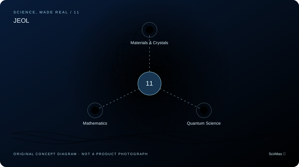
What science makes it possible The
electrons enables high spatial resolution, but lens aberration, sample thickness, beam damage, and contrast transfer collectively limit the interpretable information.
Understanding the product requires putting a single formula back into the system: input measurements, materials and boundary conditions are calculated by the model and then compared with actual data. Approximations at different scales cannot be unconditionally spliced, and relevant tools will indicate applicable conditions respectively.
Commercial applications and engineering boundaries
materials team combines image, diffraction and spectroscopy evidence to study defects, interfaces and nanostructures. The wavelength conversion on this site cannot represent the resolution of the entire machine. The
is an independent technical interpretation based on the manufacturer's public information and does not represent the manufacturer's adoption, endorsement or sponsorship of SciAtlas. The illustrations are original illustrations of the principles of this site, not photos of the actual products. Product specifications, certifications and availability are subject to the manufacturer's current information; the relevant tools only demonstrate public simplified models.
Manufacturer’s products and official information ↗Use online tools to understand related issues
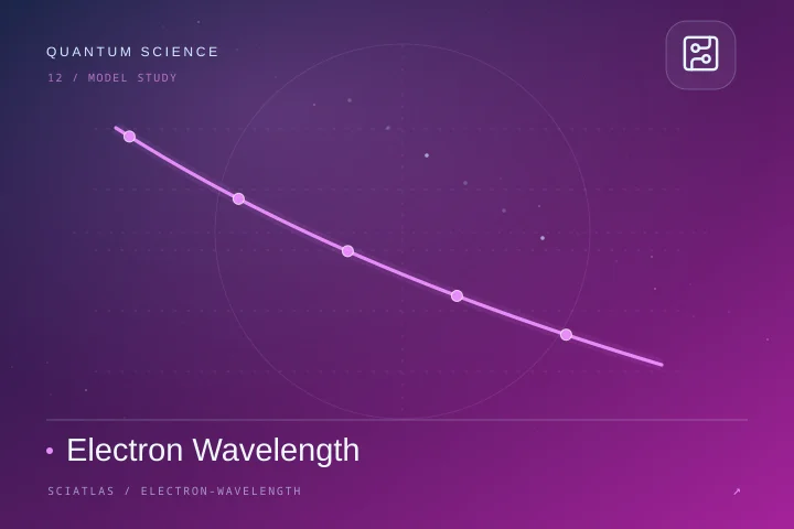
Electron Wavelength
Non-relativistic electrons accelerate from rest, high voltage requires relativistic correction.
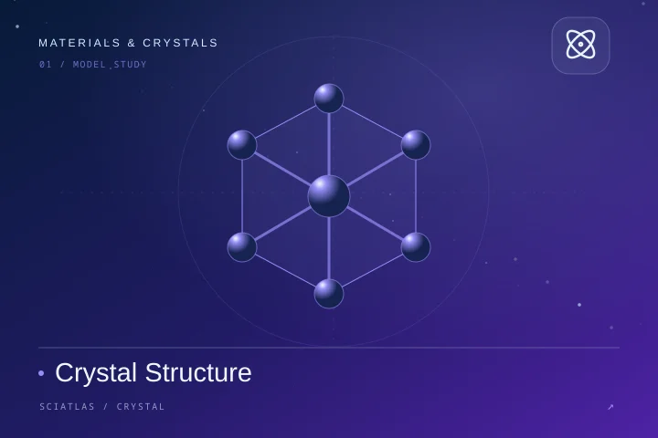
Crystal Structure
supports CIF and direct coordinate/Cartesian coordinate POSCAR; CIF expansion space group symmetry operation. Some occupancies are displayed as entered and unordered microstructure is not inferred.
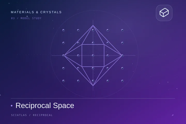
Reciprocal Space
General triclinic unit cell; the first Brillouin zone is intersected by the nearest reciprocal point half space. Diffraction only gives the geometric peak positions and does not calculate the structure factor intensity.
FFT Spectrum
Real signal, uniform sampling, rectangular window; not power spectral density.
linked model is used for independent learning and magnitude analysis and does not copy the manufacturer's proprietary software or pass product performance certification.
People and historical contributions behind the science
The following relationships distinguish theoretical and method pioneers and industry leaders; it does not mean that these figures independently invented the product on this page.
Pioneer in the development of electron microscopes, he won the 1986 Nobel Prize in Physics for his work related to electron optics and the design of the first electron microscope. The short wavelength of
William Lawrence BraggWilliam Lawrence Braggproposed the crystal diffraction relationship, developed X-ray crystal structure analysis with his father, and won the 1915 Nobel Prize in Physics.
Related Elements and Materials Basics
elemental portal is for understanding related materials or molecular basis and does not claim a complete materials list for a specific model.